
WhatsApp USA: +1 6032769350 | WhatsApp HK: +852 63697831
Global Healthcare Medical
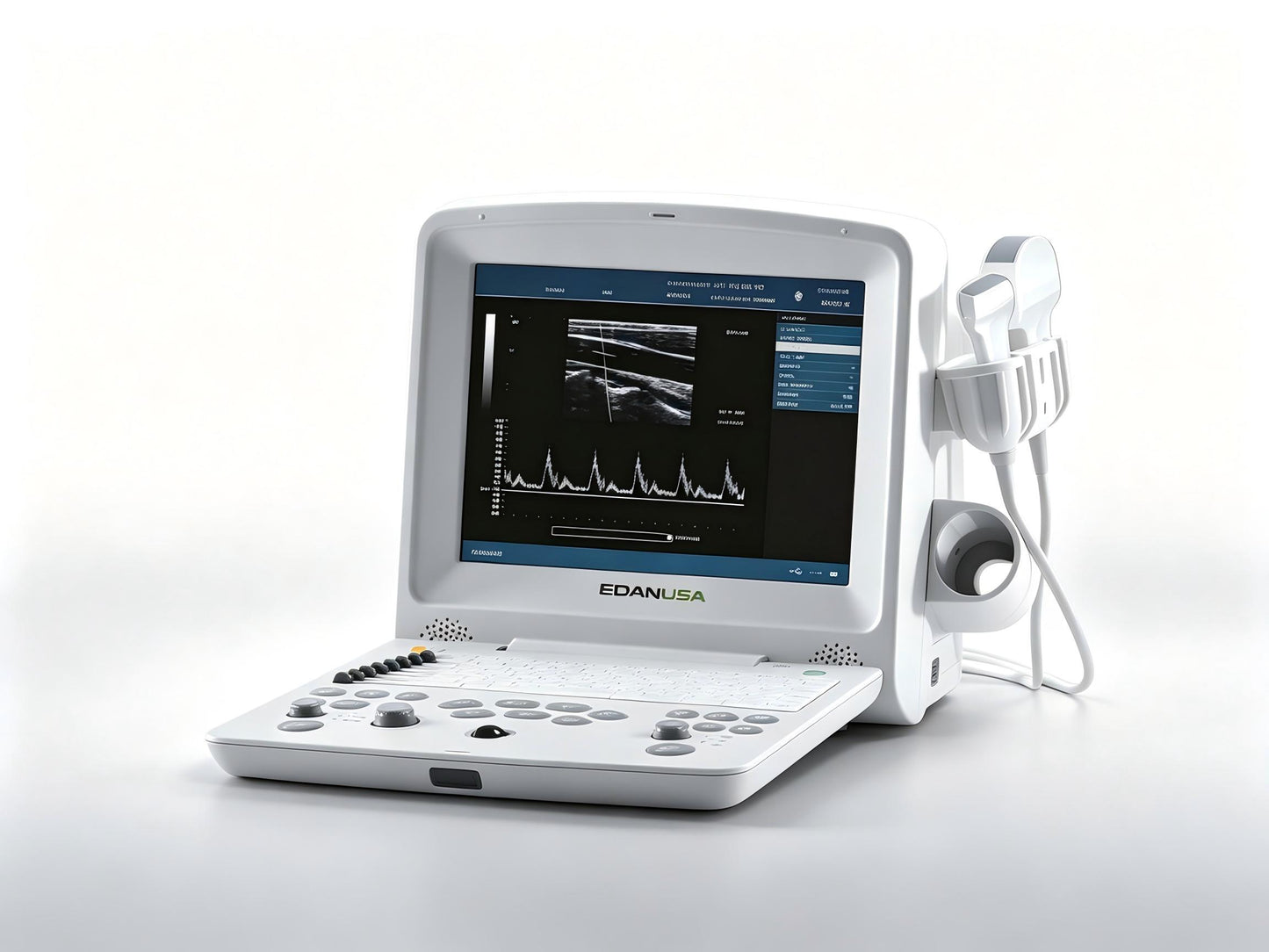
EDAN DUS 60 Digital Ultrasound System
EDAN DUS 60 Digital Ultrasound System
The EDAN DUS 60 is a portable, full‑digital diagnostic ultrasound system designed to deliver high‑quality imaging across a wide range of clinical applications. Weighing just 7.1 kg and featuring a 12.1‑inch TFT‑LCD display, the DUS 60 combines exceptional portability with robust imaging capabilities—making it an ideal solution for hospitals, clinics, and point‑of‑care settings where mobility and reliability are essential.
Equipped with two active transducer connectors and supporting frequencies from 2.0 to 10.0 MHz, the system accommodates a comprehensive selection of probes including convex, linear, micro‑convex, endovaginal, and endorectal arrays. With advanced imaging modes including B, 2B, 4B, B+M, M, and PW Doppler, the DUS 60 enables confident diagnosis across abdomen, obstetrics, gynecology, urology, small parts, musculoskeletal, and peripheral vascular applications.
Key Features & Benefits
Superior Image Quality
The DUS 60 leverages a suite of advanced imaging technologies to deliver sharp, clear diagnostic images:
-
Phase Inversion Harmonic Imaging – enhances tissue differentiation and reduces artifacts
-
Multi‑Beam Technology – improves frame rate and image consistency
-
Synthetic Receiving Aperture – optimizes lateral resolution across the entire field of view
-
Dynamic Receiving Focusing – maintains image sharpness at all depths
-
Real‑time Dynamic Aperture – adapts aperture size for optimal image quality
-
Dynamic Frequency Scanning – adjusts frequency to balance resolution and penetration
-
Dynamic Apodization – reduces side‑lobe artifacts for cleaner images
256 gray scales ensure detailed visualization of tissue structures, while multiple‑pseudo‑color imaging enhances differentiation of subtle anatomical features.
Comprehensive Imaging Modes
The DUS 60 supports a full range of imaging modes to meet diverse clinical needs:
-
B‑mode – fundamental 2D imaging
-
2B and 4B – multi‑view displays for simultaneous comparison
-
B+M – combined anatomical and motion imaging
-
M‑mode – detailed motion analysis for cardiac and fetal applications
-
PW (Pulsed Wave) Doppler – quantitative blood flow velocity assessment
Intuitive Workflow and User Convenience
Designed with the user in mind, the DUS 60 streamlines every step of the examination:
-
12.1‑inch TFT‑LCD display – delivers crisp, high‑contrast images in any lighting condition
-
Intelligent 8‑segment TGC (Time Gain Compensation) – allows precise adjustment of gain at different depths for consistently optimized images
-
Pre‑ and post‑processing capabilities – including dynamic range adjustment, frame persist, gray map selection, speckle reduction, and pseudo‑color
-
Cine loop memory – stores up to 256 frames for bidirectional review and retrospective analysis
-
Panoramic zoom – supports real‑time and freeze‑frame magnification from X1.0 to X4.0 for detailed examination of regions of interest
-
Over 130 body marks – facilitates accurate documentation and reporting
Extensive Measurement & Calculation Packages
The DUS 60 comes equipped with comprehensive measurement and calculation software for multiple specialties:
| Application | Key Measurements |
|---|---|
| General | Distance, circumference, area, volume, ratio, % stenosis, histogram, angle |
| Obstetrics | Fetal biometrics, gestational age, estimated fetal weight |
| Gynecology | Uterine and ovarian measurements |
| Small Parts | Thyroid, breast, testicular assessments |
| Orthopedics | Musculoskeletal measurements |
| Cardiology | Cardiac dimensions and function |
| Peripheral Vascular | Vessel diameter and flow assessment |
| Urology | Renal, bladder, and prostate measurements |
M‑mode measurements include distance, time, slope, and heart rate. Doppler measurements include time, heart rate, velocity, acceleration, trace, and resistive index (RI).
Portable and Durable Design
The DUS 60 is engineered for mobility without compromising performance:
-
Weight: 7.1 kg (15.7 lb) – easily transportable between departments or facilities
-
Dimensions: 330mm (L) × 220mm (W) × 320mm (H)
-
Lithium‑ion battery – supports continuous operation for up to 2 hours for uninterrupted bedside or remote examinations
-
Durable construction – built to withstand the demands of daily clinical use
Flexible Connectivity and Data Management
The DUS 60 offers comprehensive connectivity options for seamless integration into any clinical workflow:
-
504MB built‑in image storage for on‑device archiving
-
2 USB ports – for data export, software updates, and peripheral connectivity
-
Ethernet port – enables network integration and data sharing
-
Video output, S‑video output, and VGA output – for connection to external displays and recording devices
-
DICOM 3.0 compatibility (optional) – ensures seamless integration with PACS and hospital information systems
-
UMS100 workstation software (optional) – for advanced data management and reporting
Transducer Compatibility
The DUS 60 supports a wide range of transducers for maximum clinical versatility, with auto‑detection for plug‑and‑play convenience:
| Transducer | Type | Frequency Range |
|---|---|---|
| C361‑2 | Convex array | 2.5/3.5/4.5/H2.5/H2.7 MHz |
| C611‑2 | Micro‑convex array | 5.5/6.5/7.5/H4.5/H4.7 MHz |
| L743‑2 | Linear array | 6.5/7.5/8.5/H4.5/H4.7 MHz |
| L761‑2 | Linear array | 6.5/7.5/8.5/H4.5/H4.7 MHz |
| E611‑2 | Endovaginal | 5.5/6.5/7.5/H4.5/H4.7 MHz |
| E741‑2 | Endorectal | 6.5/7.5/8.5/H4.5/H4.7 MHz |
Scanning angle reaches up to 152 degrees (transducer‑dependent), and scanning depth ranges from 19 to 324 mm for comprehensive anatomical coverage.
Technical Specifications
| Specification | Detail |
|---|---|
| Imaging Modes | B, 2B, 4B, B+M, M, PW Doppler |
| Gray Scales | 256 |
| Display | 12.1‑inch TFT‑LCD |
| Transducer Frequency | 2.0 – 10.0 MHz |
| Transducer Connectors | 2 standard (with auto‑detection) |
| Scanning Angle | Up to 152° (transducer dependent) |
| Scanning Depth | 19 – 324 mm (transducer dependent) |
| Cine Loop | 256 frames, bidirectional |
| Image Storage | 504MB built‑in |
| Body Marks | >130 types |
| Power Supply | 100V – 240V, 50/60Hz |
| Battery | Lithium‑ion, up to 2 hours continuous operation |
| Dimensions (L×W×H) | 330 × 220 × 320 mm |
| Weight | 7.1 kg |
| Peripheral Ports | 2× USB, 1× Ethernet, 1× VGA, 1× Video, 1× S‑video, 1× Remote control, 1× Footswitch |
Clinical Applications
The EDAN DUS 60 is versatile enough to support a broad spectrum of clinical specialties:
-
Abdomen – liver, gallbladder, pancreas, spleen, kidney evaluation
-
Obstetrics – fetal biometry, gestational age assessment, fetal well‑being
-
Gynecology – uterine and ovarian evaluation
-
Small Parts – thyroid, breast, testicular, and superficial structure imaging
-
Musculoskeletal – conventional and superficial musculoskeletal assessment
-
Cardiology – basic cardiac evaluation with M‑mode and PW Doppler
-
Peripheral Vascular – vessel assessment and flow evaluation
-
Urology – renal, bladder, and prostate examinations
The system is applicable for adults, pregnant women, and pediatric patients in both hospital and clinic settings.
Why Choose the EDAN DUS 60?
| Clinical Challenge | DUS 60 Solution |
|---|---|
| Need for portable ultrasound | Lightweight 7.1 kg design with 2‑hour battery life—use anywhere |
| Multiple clinical applications | Comprehensive transducer range + application‑specific software packages |
| Limited workspace | Compact footprint fits any examination room |
| Image quality concerns | Harmonic imaging, multi‑beam, and dynamic focusing technologies |
| Training requirements | Intuitive user interface with 8‑segment TGC and pre‑set exam types |
| Data management | 504MB storage + DICOM 3.0 compatibility |
| Budget constraints | Cost‑effective portable system without compromising image quality |
| Department versatility | One system serves radiology, OB/GYN, urology, and emergency departments |