
WhatsApp USA: +1 6032769350 | WhatsApp HK: +852 63697831
Global Healthcare Medical
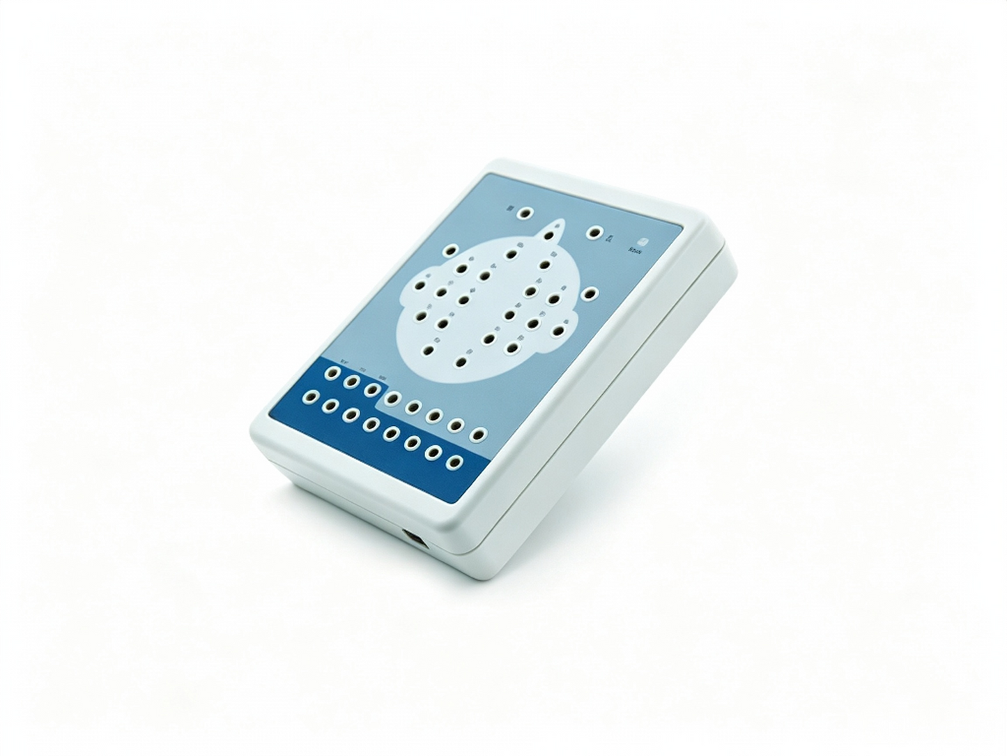
GHM-KT88 Digital Brain Electric Activity Mapping | EEG System for Neurological Diagnosis
GHM-KT88 Digital Brain Electric Activity Mapping | EEG System for Neurological Diagnosis
Digital EEG mapping system for epilepsy, brain tumors, and cerebrovascular diagnosis. 24-hour recording, FFT analysis, BEAM mapping, and PC software. Ideal for neurology clinics and hospitals.
The Global Healthcare Medical GHM-KT88 Digital Brain Electric Activity Mapping system is a comprehensive, PC‑based electroencephalography (EEG) solution designed for advanced neurological diagnosis and research. The system collects EEG signals via electrodes placed according to the international standard 10/20 system. Through integrated amplification, A/D transformation, and PC‑based auto‑analysis using Fast Fourier Transform (FFT) , the system forms electroencephalograms that display with color depth.
This product is applicable for checking such diseases as epilepsy, intracranial inflammation, cerebrovascular diseases, and brain tumors. It is an essential tool for neurologists, epileptologists, and clinical researchers seeking high‑quality EEG data acquisition and analysis.
This device is intended for professional use in clinical and research settings by trained healthcare professionals. Not a substitute for professional medical diagnosis.
Key Features
🧠 High‑Performance EEG Acquisition
The GHM-KT88 features a high‑performance bioelectricity amplifier for distilling brainwave signals with exceptional clarity. The system supports continuous recording of up to 24 hours, making it ideal for capturing episodic neurological events and conducting extended sleep studies.
🎯 Advanced Analysis & Visualization
The system features powerful automatic analysis functions that can perform power spectrum analysis and pathologic wave detection for appointed waveforms. Multiple graphs can be displayed on the same screen, including:
🔄 Flexible Playback & Review
The GHM-KT88 offers robust playback capabilities with adjustable amplitude and display speed. A special subdividing time line divides the waveform in one second into 5 parts, making it easy for clinicians to review waveforms in detail. Leads can be changed during replaying, and different types of combined leads are supported during sampling.
📋 Event Marking & Customization
EEG waveforms can be marked under different events (opening eyes, closing eyes, flashing) with different colors. User‑defined events can be added, and waveform colors for evoked events can be set freely, ensuring that waveforms at corresponding times can be rapidly found by event name during case playback.
🔬 Precision Measurement Tools
The system includes electronic frequency rulers for convenient measurement of any appointed EEG waveform. A partial enlarging window enables accurate measurement of EEG period, amplitude, and frequency, which can be adjusted according to clinical judgment.
⚙️ Fully Digital Filter System
The digital filter system can be set as required, providing different window types. Filter constants are all digital and free to enact, offering maximum flexibility for artifact removal and signal optimization.
✂️ EEG Signal Clipping & Analysis
The system features an EEG signal clipping function that allows users to analyze and store any section of EEG wave, and select several waveform segments for automatically analyzing and distilling to different parameters.
🔌 USB Interface & Flash Stimulator
The system uses a USB interface for data transmission. A multifunctional flash stimulator connects via USB, with flashing that can be controlled manually or automatically. A flash stimulation scheme can be set and performed during the sampling process.
🛡️ Premium Safety Features
The GHM-KT88 incorporates a professional isolation transformer, dual power supply isolation system, and optoelectronic data transmission to ensure patient and device safety. The system also features an integrated full‑automatic calibration system.
💾 Comprehensive Case Management
The system features a perfect case management function, providing multiple means for research and quick statistical information. Cases can be exported and imported conveniently, with storage options including MO or CD‑RW disks. Case files can be transformed into EDF and BDF data formats, facilitating data exchange, academic exchange, and further analysis.
📄 Integrated Reporting
The system generates integrative image and character reports that can be edited in mode and switched to Word documents. This flexibility streamlines clinical documentation and research publication.
📷 Optional Video & SpO₂ Functions
Optional functions include video recording via USB camera with flexible playback and SpO₂ monitoring. (Not all optional functions are available in all regions.)
Specifications
Applications
The GHM-KT88 Digital Brain Electric Activity Mapping system is suitable for:
-
Neurology Departments – Diagnosis and monitoring of epilepsy, intracranial inflammation, cerebrovascular diseases, and brain tumors
-
Epilepsy Clinics – Comprehensive EEG analysis for seizure detection and classification
-
Sleep Medicine – Extended 24‑hour recordings for sleep disorder assessment
-
Research Institutions – Advanced brain activity mapping and academic research
-
Intensive Care Units (ICU) – Continuous brain monitoring in critical care settings
-
Neurosurgery Departments – Preoperative and postoperative brain function assessment
Why Choose the GHM-KT88?
FAQ
Q: What is the GHM-KT88 Digital Brain Electric Activity Mapping system?
A: The GHM-KT88 is a comprehensive EEG system that collects brain signals via electrodes, processes them through amplification and A/D conversion, and uses PC‑based auto‑analysis with FFT to generate color‑coded brain activity maps.
Q: What electrode placement method does the system use?
A: The system uses the international standard 10/20 electrode placement method.
Q: How long can the system record continuously?
A: The GHM-KT88 supports continuous recording of up to 24 hours.
Q: What analysis functions are available?
A: The system provides power spectrum analysis, pathologic wave detection, BEAM mapping, numerical BEAM, compressed spectrum graphs, and trend graphs.
Q: Can I mark events during recording?
A: Yes. EEG waveforms can be marked for events such as opening eyes, closing eyes, and flashing with different colors. User‑defined events can also be added.
Q: What data formats are supported for export?
A: Case files can be transformed into EDF and BDF data formats for data exchange and further analysis.
Q: What safety features does the system have?
A: The system features a professional isolation transformer, dual power supply isolation system, and optoelectronic data transmission for enhanced safety.
Q: What optional functions are available?
A: Optional functions include video recording via USB camera and SpO₂ monitoring. (Not all optional functions are available in all regions.)
Q: What clinical conditions is the system suitable for?
A: The system is applicable for checking epilepsy, intracranial inflammation, cerebrovascular diseases, and brain tumors.