

WhatsApp USA: +1 6032769350 | WhatsApp HK: +852 63697831
Global Healthcare Medical
SonoScape P15 Trolley Color Doppler Ultrasound System
SonoScape P15 Trolley Color Doppler Ultrasound System
The SonoScape P15 is a trolley-based color Doppler ultrasound system engineered to enhance ultrasound exams across a broad range of clinical specialties—from gynecology and cardiology to internal medicine, abdominal, and superficial imaging. It inherits the Wi-Sono high-end platform and combines it with advanced probe technology and a suite of intelligent image-processing tools, delivering high-resolution images with excellent penetration depth for precise and reliable assessment.
The P15 is positioned as a cost-effective, simplified console with an intuitive user interface and multiple intelligent functions, designed for clinicians who need diagnostic confidence without a complex or oversized cart footprint. The system's broad bandwidth platform enables better information collection, providing higher resolution at depth and enhanced image quality for accurate diagnosis across different patient body habitus.
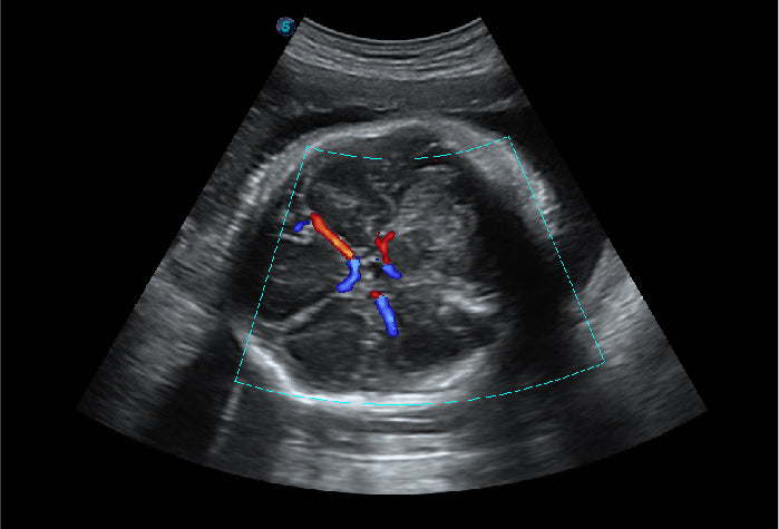
The imaging chain incorporates Spatial Compound Imaging for optimal contrast resolution, speckle reduction, and border detection—making the platform ideal for superficial and abdominal imaging with better clarity and improved continuity of structures. μ-Scan+ further reduces image noise, improves signal strength, and enhances overall visualization. Dynamic Color technology builds on existing color Doppler methods to enable clearer capture of color flow and detailed visualization of even tiny veins with lower velocities.
The P15 supports B-mode (2B & 4B), M-mode, Color Doppler Flow Imaging, Pulsed Wave Doppler, High Pulse Repetition Frequency, Tissue Harmonic Imaging, Tissue Specific Imaging, and Pulse Inversion Harmonic Imaging, alongside 3D/4D imaging, Real-time Panoramic, Tissue Doppler Imaging (TDI), and Auto IMT. The system is configured with a 21.5-inch Full HD LED monitor with a wide viewing angle and a 13.3-inch touchscreen with adjustable tilt for ergonomic operation.
Regulatory status: The P15 platform is documented with CE marking and ISO 13485 certification in distributor materials. No FDA 510(k) clearance specific to the P15 model name was identified in the public FDA database during preparation of this page; the P15 Elite/Exp variants are listed in the FDA Device Listing under Product Code IYN (K221140) as part of the P20 Elite Series family, but the base P15 designation should be confirmed against local registration records before purchase. No independent peer-reviewed clinical validation study specific to the P15 was identified in the public literature during preparation of this page. Availability, labeling, and cleared claims vary by market.
Key Features & Benefits
Broad Bandwidth Platform with Advanced Probe Technology
The broad bandwidth platform and advanced probe technology enable better information collection, delivering higher resolution at depth and enhanced image quality for accurate diagnosis across superficial, abdominal, and deeper tissue layers. The system is compatible with a wide range of ultrasound probes for various specialties, supporting flexible clinical coverage within a single cart configuration.
Spatial Compound Imaging and μ-Scan+ for Image Clarity
Spatial Compound Imaging uses multiple lines of sight to achieve optimal contrast resolution, speckle reduction, and border detection—making the P15 ideal for superficial and abdominal imaging with better clarity and improved continuity of anatomical structures. μ-Scan+ is a new-generation imaging technology that reduces noise, improves signal strength, and enhances visualization for clearer tissue definition and lesion conspicuity.
Dynamic Color for Fine Vascular Visualization
Dynamic Color improves upon existing color Doppler technologies to enable clearer capture of color flow and detailed visualization of even tiny veins with lower velocities—supporting hemodynamic assessment in fine vascular structures where conventional color Doppler may struggle to resolve slow flow.
Real-Time Panoramic and 3D/4D Imaging
Real-time Panoramic acquires an extended field of view for large organs or long vessels, making measurement easier and enhancing diagnostic efficiency. The acquisition is performed in real time, and any mistake can be easily back-tracked and corrected without interrupting the scan. 3D/4D imaging delivers outstanding volume performance with speed and convenience, supporting detailed volumetric visualization of anatomical structures.
Tissue Doppler Imaging and Auto IMT
Tissue Doppler Imaging (TDI) allows clinicians to quantitatively evaluate local myocardial movements and functions, supporting analysis and comparison of the motions of different parts of the heart. Auto IMT provides quick measurement of intima-media vessel thickness, ensuring good reproducibility and high diagnostic efficiency in cardiovascular examinations.
Ergonomic Console with 21.5-Inch Full HD Monitor and 13.3-Inch Touchscreen
The system features a 21.5-inch Full HD LED monitor with a wide viewing angle for brilliant image display, alongside a 13.3-inch touchscreen with adjustable tilt for ergonomic operation. The clear control panel includes an extendable, illuminated keyboard and an intuitive layout with logically arranged controls. Five probe ports—four active and one in park position—support quick probe switching during multi-specialty examinations. Four smooth-rolling casters with locking brakes provide maximum mobility in clinical settings.
Connectivity: DICOM, USB, HDMI, and Wi-Fi
The P15 supports DICOM 3.0 (Store, C-Store, Worklist, MPPS, Print, SR, Q&R), USB, HDMI, and Wi-Fi for seamless data transfer and PACS/HIS integration where configured. Generous internal storage supports patient image and data retention.
Technical Specifications
Why Choose the SonoScape P15?