

WhatsApp USA: +1 6032769350 | WhatsApp HK: +852 63697831
Global Healthcare Medical
SonoScape P50 Trolley Color Doppler Ultrasound System
SonoScape P50 Trolley Color Doppler Ultrasound System
The SonoScape P50 is a slim, trolley-based color Doppler ultrasound system engineered for shared-service clinical imaging across fetal, abdominal, pediatric, small-organ, cephalic, trans-rectal, trans-vaginal, peripheral vascular, cerebral vascular, musculoskeletal, cardiac, trans-esophageal, laparoscopic, OB/GYN, and urology applications. It combines single-crystal wide-band transducers, μ-Scan+ speckle reduction, and a high-sensitivity touchscreen interface in a compact chassis designed for tight clinical spaces—where a full-size cart may not fit but comprehensive imaging capability is still required.
The P50 Series received FDA 510(k) clearance under K170999 (July 12, 2017) as a Class II ultrasonic pulsed Doppler imaging system (Product Code IYN, 21 CFR 892.1550) , following a Track 3 traditional review. The FDA summary states “No clinical testing was required” ; substantial equivalence was established through bench testing covering electrical, mechanical, thermal, electromagnetic compatibility, biocompatibility, and acoustic output, conforming to relevant IEC, ISO, AIUM, and NEMA standards. The P50 Elite is also registered with the Medical Device Authority of Malaysia (MDA) under GB9407422-113585 (valid 15 December 2022 – 14 December 2027) as part of the P40/P50/S50 Elite Series. No independent peer-reviewed clinical validation study specific to the P50 was identified in the public literature during preparation of this page.
The imaging chain is anchored by single-crystal wide-band probes that improve signal-to-noise ratio and acquire images with superior sensitivity and resolution in both near and far fields, with a 1.0–25.0 MHz frequency range supporting linear, convex, and phased array probes across superficial through deep applications. μ-Scan+ reduces noise and improves signal strength and visualization, while Dynamic Color enhances color Doppler capture for tiny veins with lower velocities. C-xlasto Imaging visualizes tissue stiffness variations for breast, thyroid, and musculoskeletal assessment, and Contrast Imaging uses infra-harmonic and second-harmonic signals with Dynamic Acoustic Control to support lesion perfusion observation at reduced agent doses.
Key Features & Benefits
Single-Crystal Wide-Band Probes
The P50 is compatible with SonoScape‘s single-crystal probes, which greatly improve signal ratio and provide superior sensitivity and resolution for both the near and far fields—supporting clear 2D clinical images and Doppler sensitivity for critical cardiac performance assessment. The wide-band design covers a 1.0–25.0 MHz range across linear, convex, and phased array configurations, enabling a single platform to serve superficial, abdominal, and cardiac workflows within the physical limits of the installed probe set.
μ-Scan+ and Dynamic Color
μ-Scan+ is a next-generation imaging technology that reduces noise, improves signal strength, and improves visualization for clearer tissue definition and lesion conspicuity. Dynamic Color improves upon existing color Doppler technologies to capture color flow clearly and visualize even tiny veins with lower velocities—supporting hemodynamic assessment in fine vascular structures.
C-xlasto Imaging and Contrast Imaging with Dynamic Acoustic Control
C-xlasto Imaging detects and visualizes differences in tissue responses through elastography algorithms in real time, supporting breast, thyroid, and musculoskeletal assessment; SonoScape’s transvaginal and bi-plane probes extend elastography applications into gynecology and urology. Contrast Imaging on the P50 uses infra-harmonic and second-harmonic signals to improve image resolution and deep penetration, while Dynamic Acoustic Control technology effectively controls acoustic pressure for the contrast agent, decreasing the required agent dose, assuring uniform image quality, and supporting longer contrast duration and better delayed-phase lesion perfusion observation.
OB/GYN Tools: S-Live Silhouette, Pelvic Floor 4D, and Auto NT
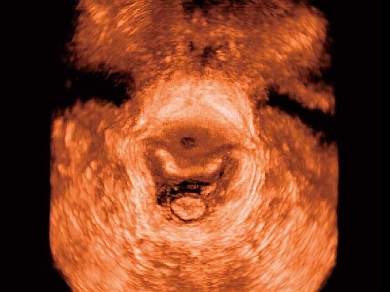
The P50 includes a transparent 3D anatomical rendering (S-Live Silhouette) that depicts fetal intracorporeal anatomical structure in a way conventional ultrasound cannot. Pelvic Floor 4D works with transvaginal probes for trans-perineal assessment of anterior compartment impact after vaginal delivery, supporting evaluation of pelvic organ prolapse and muscle integrity. Auto NT automatically traces and calculates nuchal translucency thickness for faster evaluation, and Auto Face removes masking layers in front of the fetal face for a clearer view. AVC Follicle automatically identifies follicle count and individual volumes.
Cardiology Tools: TDI, Stress Echo, Auto IMT, and Auto EF
Tissue Doppler Imaging (TDI) allows clinicians to quantitatively evaluate local myocardial movements and functions, supporting analysis and comparison of different parts of the heart. Stress Echo combines 2D echocardiography with physical, pharmacological, or electrical stress, providing configurable template editing, wall motion scoring, and stress echo reporting. Auto IMT automatically traces and calculates carotid vessel thickness with instant Mean and Max indices at a single button press. Auto EF provides an intelligent endocardial trace function with 19 adjustable points for rapid 2D EF and volume assessment.
Slim Trolley Design with 21.5-Inch LED Monitor and 13.3-Inch Touchscreen
The P50 is a slim and unobtrusive trolley system comfortable in tight, congested spaces with little room to work—providing everything needed for a comfortable examination in a small space for both clinician and patient. The configuration includes a 21.5-inch high-resolution LED color monitor, a 13.3-inch high-resolution touchscreen, a simplified control panel, five transducer connectors (including one CW transducer connector), an endocavity probe holder, gel warmer, built-in battery, and wireless Wi-Fi connection. 500 GB hard disk, DVD-RW, and USB 3.0 support data storage and export, and DICOM 3.0 (Store/C-Store/Worklist/MPPS/Print/SR/Q&R) supports PACS and HIS integration where configured.
Technical Specifications
| Specification | Detail |
|---|---|
| Device Type | Trolley-based color Doppler ultrasound system |
| Regulatory Status | FDA 510(k) K170999 (Jul 12, 2017) — P50 Series Digital Color Doppler Ultrasound System; Class II, Product Code IYN, 21 CFR 892.1550; Track 3 traditional review; “No clinical testing was required” per FDA summary; MDA Malaysia: GB9407422-113585 (P40/P50/S50 Elite Series; valid 15 Dec 2022 – 14 Dec 2027); no independent peer-reviewed clinical validation study specific to P50 identified |
| Frequency Range | 1.0–25.0 MHz |
| Probe Types | Linear array, convex array, phased array; single-crystal wide-band probes; transvaginal and bi-plane probes for elastography extensions |
| Imaging Modes | B-Mode (including Tissue Harmonic Imaging), M-Mode, TDI, Color-Flow Doppler, Pulsed Wave Doppler, Continuous Wave Doppler, Power Doppler, Elastography, 3D/4D, combined modes |
| Image Processing | μ-Scan+ (noise reduction, signal strength, visualization); Dynamic Color (tiny vein/low-velocity flow capture); C-xlasto Imaging (tissue stiffness); Contrast Imaging with Dynamic Acoustic Control |
| Clinical Tools | S-Live Silhouette, Pelvic Floor 4D, Auto NT, Auto Face, AVC Follicle, S-Guide, TDI, Stress Echo, Auto IMT, Auto EF, VIS-Needle |
| Display | 21.5-inch high-resolution LED color monitor; 13.3-inch high-resolution touchscreen |
| Probe Connectors | Five transducer connectors (including one CW connector); endocavity probe holder |
| Storage & Connectivity | 500 GB hard disk, DVD-RW, USB 3.0; DICOM 3.0 (Store/C-Store/Worklist/MPPS/Print/SR/Q&R); wireless Wi-Fi |
| Power | Built-in battery; power assistant battery operation |
| Ergonomics | Slim trolley design, simplified control panel, gel warmer, rotatable/height-adjustable control panel |
| Clinical Applications | Fetal, Abdominal, Pediatric, Small Organ, Cephalic, Trans-rectal, Trans-vaginal, Peripheral Vascular, Cerebral Vascular, Musculoskeletal, Cardiac, Trans-esophageal, Laparoscopic, OB/GYN, Urology |
| Intended Use | General-purpose ultrasonic imaging for evaluation of the clinical applications listed above by a qualified physician |
Why Choose the SonoScape P50?
| Clinical or Operational Challenge | P50 Solution |
|---|---|
| Need a full-featured ultrasound that fits in tight clinical spaces | Slim and unobtrusive trolley design for congested rooms; 21.5-inch LED monitor and 13.3-inch touchscreen |
| Need high image clarity and sensitivity for abdominal, cardiac, and vascular imaging | Single-crystal wide-band probes for improved signal-to-noise ratio, near/far field sensitivity, and resolution |
| Need tissue stiffness assessment for breast, thyroid, and MSK lesions | C-xlasto Imaging with real-time elastography; transvaginal and bi-plane probe extensions |
| Need contrast-enhanced visualization with reduced agent dose | Contrast Imaging with Dynamic Acoustic Control and infra-harmonic/second-harmonic signals |
| Need OB/GYN tools for fetal anomaly and pelvic floor assessment | S-Live Silhouette, Pelvic Floor 4D, Auto NT, Auto Face, AVC Follicle, S-Guide |
| Need cardiac functional assessment tools | TDI, Stress Echo, Auto IMT, Auto EF, and CW Doppler connector |
| Need large storage, DICOM connectivity, and battery backup | 500 GB HDD, DICOM 3.0, wireless Wi-Fi, built-in battery |
| Need FDA-listed equipment with documented bench testing evidence | FDA K170999 covering the P50 Series; bench testing only, no clinical data required |