

WhatsApp USA: +1 6032769350 | WhatsApp HK: +852 63697831
Global Healthcare Medical
SonoScape P70i Trolley Color Doppler Ultrasound System
SonoScape P70i Trolley Color Doppler Ultrasound System
The SonoScape P70i is a premium trolley-based color Doppler ultrasound system engineered for women's health, obstetrics, gynecology, and shared-service imaging. It is built on SonoScape's C-Field+™ architecture, a platform designed to optimize signal-to-noise ratio, spatial and contrast resolution, and frame rate while integrating accelerated computing for efficient clinical processing. The system is positioned as a shared-service platform covering abdomen, vascular, cardiology, OB/GYN, small parts, MSK, and interventional applications, with a particular emphasis on maternal and gynecologic imaging.
The P70i is listed in the FDA Establishment Registration & Device Listing under the proprietary name “P70i Digital Color Doppler Ultrasound System” , with Product Code IYO (System, Imaging, Pulsed Echo, Ultrasonic), Class 2, Regulation 892.1560, and Premarket Submission Number K222596 cited in the listing. The K222596 clearance covers the S90 Exp Series Digital Color Doppler Ultrasound System, which includes the P70i, S70i, P80 Elite, S80 Elite, S100 Exp, P90i and related models as a shared-family submission. No independent peer-reviewed clinical validation study specific to the P70i was identified in the public literature during preparation of this page. Availability, labeling, and cleared claims vary by market.
The imaging chain is anchored by a premium transducer portfolio—convex array, volume probe, linear array, phased array, and specialty probes—designed for women's health examinations and shared-service applications. Advanced hemodynamic tools include FHR for microvascular structure visualization, Bright Flow for 3D-like color Doppler flow without a volume transducer, and Micro F for separating subtle flow signals from tissue motion.
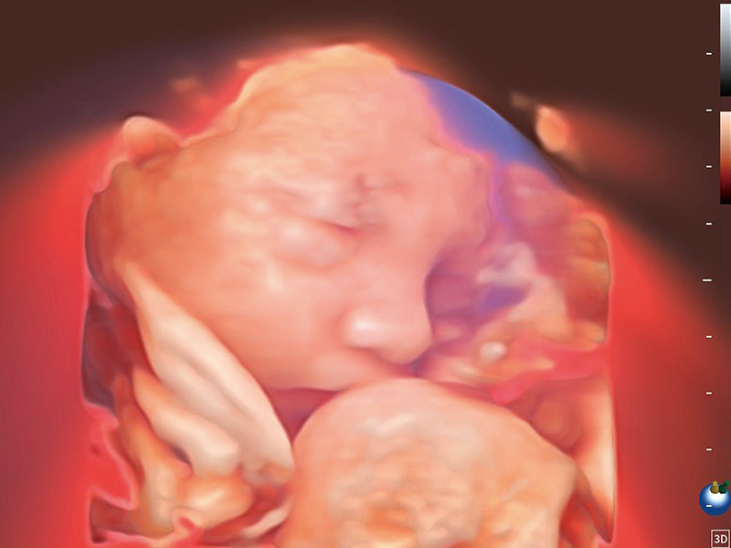
The P70i is positioned for women's wellness across the care continuum: reproductive health, early pregnancy screening, second and third trimester examination, fetal heart examination, pelvic floor examination, and gynecology care. Volume assessment and AI-assisted tools include S-Live Studio for realistic skin visualization, S-Fetus 5.0 for automatic fetal plane recognition and biometry (29 sections, 13 measurements), AVC Follicle for follicle analysis with color-coded sorting, HyCoSy with SPI for fallopian tube patency assessment, S-PF (3D) for AI-driven levator hiatus evaluation, and S-ERA for automatic endometrial segmentation and volume quantification.
Workflow assistants include Probe Auto Wakeup for automatic transducer activation, SonoSynch for real-time remote consultation, SonoDrop for wireless image and report transfer, SonoAssistant for automated protocols, SonoHelp for on-screen tutorials, and SonoTransfer for Windows-based data sharing.
Key Features & Benefits
C-Field+™ Architecture for Imaging and Clinical Efficiency
The C-Field+™ platform is designed to optimize signal-to-noise ratio, spatial and contrast resolution, and frame rate, while accelerated computing supports boosted processing for clinical workflows. The architecture is intended to deliver high-quality imaging across different body habitus and clinical applications, within the physical limits of the installed probe set.
Sophisticated Volume Assessment: S-Live Studio, S-Fetus 5.0, S-PF (3D), and S-ERA
S-Live Studio simulates light propagation on the skin's surface to deliver realistic real-time skin visualization and enhance 3D image experience for fetal assessment. S-Fetus 5.0 intelligently identifies 29 standard fetal sections and automatically obtains 13 biometric measurements with a single click. S-PF (3D) supports AI-driven Levator Hiatus evaluation for pelvic floor assessment. S-ERA enables automatic segmentation of the endometrium and volume quantification for endometrial receptivity analysis.
AVC Follicle and HyCoSy with SPI for Reproductive Assessment
AVC Follicle supports efficient follicle analysis with follicles sorted by size and rendered in different colors for intuitive visualization. HyCoSy with SPI provides sensitive contrast-timing visualization for assessment of the uterine cavity, fallopian tubes, and ovarian anatomy, supporting fallopian tube patency evaluation in infertility assessment.
Hemodynamic Tools: FHR, Bright Flow, and Micro F
FHR enables observation of microvascular structure with enhanced sensitivity. Bright Flow offers 3D-like color Doppler flow visualization without requiring a volume transducer, strengthening vessel wall definition. Micro F efficiently distinguishes subtle blood flow signals from overlaying tissue motion, enabling heightened sensitivity and spatial resolution in hemodynamic depiction. These are imaging capabilities—clinical value depends on proper probe selection and clinician review.
Versatile Workflow Assistants for Daily Efficiency
Probe Auto Wakeup uses a motion sensor for automatic transducer activation with adjustable sensitivity. SonoSynch enables real-time diagnostic interface and camera sharing for multi-device remote consultation. SonoDrop supports wireless onsite transmission of images, videos, and PDF reports. SonoAssistant helps standardize exams and reduce keystrokes and exam time. SonoHelp provides interactive tutorials for probe placement and standard examples. SonoTransfer sends PDF reports, images, and cine data to a shared Windows folder.
Premium Transducer Portfolio and Shared-Service Capability
The P70i supports a range of specialized transducers—convex array, volume probe, linear array, phased array, and specialty probes—covering women's health examinations alongside abdominal, vascular, MSK, and small-parts applications. The system is positioned as a shared-service ultrasound platform for multi-department imaging programs, supporting efficient probe switching and workflow flexibility across clinical specialties.
Technical Specifications
Why Choose the SonoScape P70i?