
WhatsApp USA: +1 6032769350 | WhatsApp HK: +852 63697831
Global Healthcare Medical
SonoScape S12 Trolley Color Doppler Ultrasound System
SonoScape S12 Trolley Color Doppler Ultrasound System
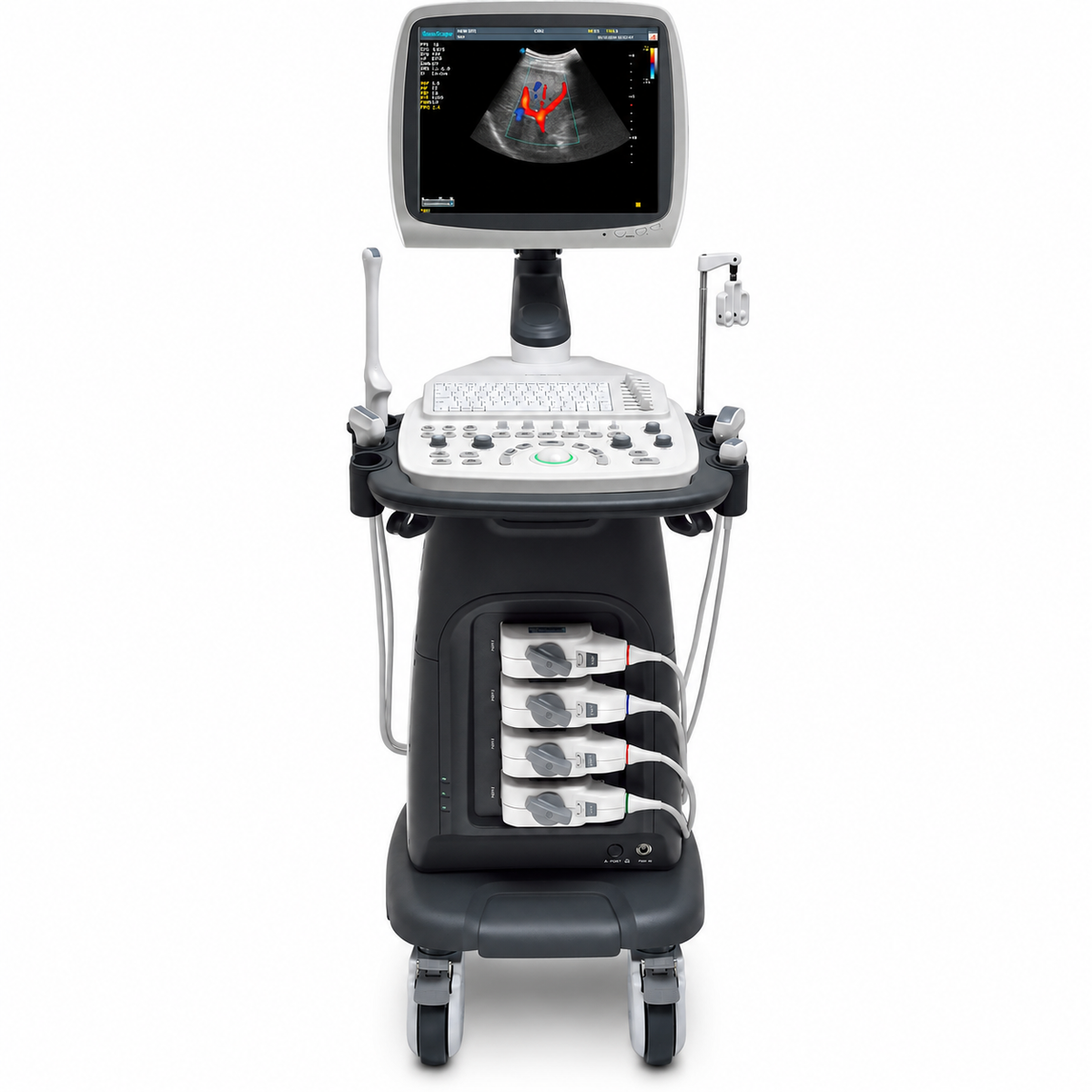
The SonoScape S12 is a compact trolley-based color Doppler ultrasound system engineered around women‘s health imaging—from routine gynecologic examinations to 2D and 3D/4D fetal assessment—while remaining adaptable to abdominal, pediatric, small-organ, vascular, musculoskeletal, and cardiac applications. It pairs a 15-inch high-definition LED monitor with an articulating arm and four active universal transducer sockets with a built-in battery, supporting flexible operation across examination rooms and bedside settings.
The S12 is FDA-cleared under 510(k) K152396 as a Class II ultrasonic pulsed echo imaging system (Product Code IYO, 21 CFR 892.1560) , following a Traditional review. A prior clearance under K142474 covers the S12 platform, and the S12 Series includes the S12EXP, S12PRO, S12, S11PLUS, M12, S10, S11S, and S12S configurations. The FDA summary for K152396 states the device is substantially equivalent to the previously cleared S12 (K142474) , with performance assessed through conformance to applicable medical device standards and design specifications—not through independent clinical trials establishing diagnostic accuracy. No independent peer-reviewed clinical validation study specific to the S12 was identified in the public literature during preparation of this page. Distributor documentation references CE marking and ISO 13485 certification for the platform.
The imaging chain supports B-Mode (including Tissue Harmonic Imaging), M-Mode, TDI, Color-Flow Doppler, Pulsed Wave Doppler, Continuous Wave Doppler, Power Doppler, Elastography, and 3D/4D imaging, with a frequency range of 1.0–15.0 MHz across linear, convex, and phased array probes. μ-Scan 2D speckle reduction, Pulse Inversion Harmonic Imaging (PIH) , Real-time Panoramic Imaging, Trapezoid Imaging, Auto-IMT, and Triplex processing are referenced in the platform’s feature set, alongside DICOM 3.0, AVI/JPG, USB 2.0, HDD, DVD, and PDF report output.
Key Features & Benefits
15-Inch HD LED Monitor with Articulating Arm and Four Active Transducer Sockets
The 15-inch high-definition LED monitor is mounted on an articulating arm for flexible positioning, supporting comfortable viewing across different scanning positions. Four active universal transducer sockets allow multiple probes to remain connected and switchable during mixed-specialty workflows, supporting efficient transitions between abdominal, endovaginal, and cardiac examinations. The system’s compact trolley design and built-in battery support mobility between rooms and bedside use.
Women‘s Health Focus: Superior Endovaginal and Fetal Imaging
The S12 is positioned as a preferred compact platform for women’s health imaging challenges—offering advanced solutions for every stage of pregnancy and gynecologic examination. The system is designed for superior endovaginal imaging and best-in-class fetal imaging in both 2D and 3D/4D acquisition. The volumetric convex array VC6-2 and endocavity 6V1 probes extend the platform into obstetric and gynecologic applications, with real-time 3D (4D) available as an optional module.
Pulse Inversion Harmonic Imaging (PIH) and μ-Scan Speckle Reduction
Pulse Inversion Harmonic Imaging fully preserves harmonic wave signal and restores authentic acoustic information, boosting resolution and reducing noise for clearer visualization. μ-Scan is a 2D speckle reduction technology that reduces noise, improves boundary signal, and elevates image uniformity for consistent tissue definition across different scanning conditions. Real-time Panoramic Imaging and Trapezoid Imaging further extend the field of view for large organs or lesions, supporting diagnosis and measurement during the scan.
Broad Probe Portfolio: Linear, Convex, Endocavitary, Phased Array, Intraoperative, and Volumetric
The S12 supports a comprehensive selection of probes—linear, convex, micro-convex, endocavity, phased array, intraoperative, bi-plane, and volumetric—covering 1.0–15.0 MHz across 128-element linear L741 (vascular, small parts, MSK), 128-element convex C344 (abdominal, obstetrics, gynecology), 72-element convex C322 (abdominal biopsy), 128-element endocavity 6V1 (gynecology, obstetrics, urology), 64-element phased array 2P1 and 5P1 (cardiac, transcranial, pediatric), 96-element linear 10I2 (intra-operative), and volumetric convex VC6-2 (obstetrics, abdominal) configurations. Needle guide brackets are referenced for ultrasound-guided procedures.
Auto-IMT, ECG Module, and Triplex Imaging
Auto-IMT provides automated carotid intima-media thickness measurement for vascular risk assessment. An ECG module is included as standard for cardiac timing applications, and Triplex imaging supports simultaneous 2D, color, and spectral Doppler display for comprehensive hemodynamic assessment. M-Tuning provides one-button image optimization for quick parameter adjustment across B, CFM, and PW modes.
500 GB Storage and DICOM 3.0 Connectivity
The system includes 500 GB hard-disk storage (with 1 TB upgrade referenced in distributor documentation), alongside DVD-RW, USB 2.0, and DICOM 3.0 (Transmission/Worklist/Print/Storage Commitment/MPPS) for PACS and hospital information system integration where configured. AVI/JPG and PDF report output support data export and documentation in routine clinical workflows.
Built-In Battery and Compact Trolley Design
The built-in battery supports operation during power interruption or intra-facility transport, supporting bedside and mobile use where mains power is not continuously available. The compact and agile trolley design with four smooth-rolling casters supports movement between examination rooms and departments, and the user-oriented multinational language input keyboard supports efficient patient information entry.
Technical Specifications
| Specification | Detail |
|---|---|
| Device Type | Trolley-based color Doppler ultrasound system |
| Regulatory Status | FDA 510(k) K152396 — S12 Digital Color Doppler Ultrasound System; Class II, Product Code IYO, 21 CFR 892.1560; prior clearance K142474 covers the S12 platform; S12 Series includes S12EXP, S12PRO, S12, S11PLUS, M12, S10, S11S, S12S; CE marking and ISO 13485 referenced in distributor documentation; no independent peer-reviewed clinical validation study specific to S12 identified |
| Display | 15-inch high-definition LED monitor with articulating arm |
| Transducer Sockets | Four active universal transducer sockets |
| Frequency Range | 1.0–15.0 MHz |
| Probe Types | Linear array, convex array, micro-convex, endocavity, phased array, intraoperative, bi-plane, volumetric; standard probes include L741 (4–16 MHz), C344 (2–6.8 MHz); optional C322, 6V1, 2P1, 5P1, 10I2, VC6-2 |
| Imaging Modes | B-Mode (including Tissue Harmonic Imaging), M-Mode, TDI, Color-Flow Doppler, Pulsed Wave Doppler, Continuous Wave Doppler, Power Doppler, Elastography, 3D/4D, combined modes |
| Image Processing | μ-Scan 2D speckle reduction; Pulse Inversion Harmonic Imaging (PIH); Real-time Panoramic Imaging; Trapezoid Imaging; Auto-IMT; Triplex; M-Tuning |
| Clinical Tools | Auto-IMT; ECG module (standard); 3D/4D (optional); Real-time 3D (4D) |
| Storage & Connectivity | 500 GB hard disk (1 TB upgrade referenced); DVD-RW; USB 2.0; DICOM 3.0 (Transmission/Worklist/Print/Storage Commitment/MPPS); AVI/JPG; PDF report |
| Battery | Built-in battery |
| Dimensions / Weight | Weight referenced as 130 kg in some distributor documentation (configuration-dependent; confirm with supplier) |
| Clinical Applications | Fetal, Abdominal, Pediatric, Small Organ (breast, testes, thyroid), Cephalic (neonatal and adult), Transrectal, Trans-vaginal, Peripheral Vascular, Musculoskeletal (Conventional and Superficial), Cardiac (neonatal and adult), OB/Gyn, Urology |
| Intended Use | General-purpose ultrasonic imaging for evaluation of the clinical applications listed above by a qualified physician |
Why Choose the SonoScape S12?
| Clinical or Operational Challenge | S12 Solution |
|---|---|
| Need a compact women’s health ultrasound with strong endovaginal and fetal imaging | 15-inch HD monitor, articulating arm, four active sockets; endocavity 6V1 and volumetric VC6-2 probes; 2D and 3D/4D acquisition |
| Need clear harmonic imaging with speckle reduction | Pulse Inversion Harmonic Imaging for resolution and noise reduction; μ-Scan for 2D speckle reduction and image uniformity |
| Need extended field-of-view for large organs or lesions | Real-time Panoramic Imaging and Trapezoid Imaging |
| Need a broad probe portfolio for multi-specialty workflows | Linear, convex, micro-convex, endocavity, phased array, intraoperative, bi-plane, and volumetric probes |
| Need automated vascular measurement and cardiac timing | Auto-IMT and ECG module (standard) |
| Need DICOM connectivity and large storage | DICOM 3.0, 500 GB HDD, DVD-RW, USB 2.0, AVI/JPG, PDF report |
| Need mobility with power backup | Built-in battery and compact trolley design with four casters |
| Need FDA-cleared equipment with documented family clearance | FDA K152396 and K142474 covering the S12 platform; CE marking and ISO 13485 referenced in distributor documentation |