
WhatsApp USA: +1 6032769350 | WhatsApp HK: +852 63697831
Global Healthcare Medical
SonoScape S22 Trolley Color Doppler Ultrasound System
SonoScape S22 Trolley Color Doppler Ultrasound System
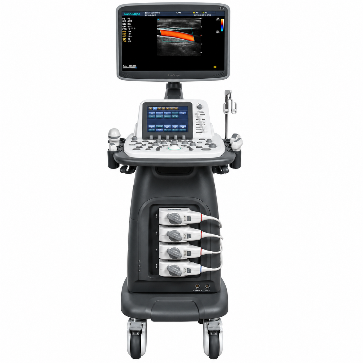
The SonoScape S22 is a shared-service trolley ultrasound system engineered to bring excellence to any clinical setting—from the emergency department, ICU, and operating room to routine general imaging. It combines a slim and elegant package with built-in battery mobility, a comprehensive probe portfolio, and premium application technologies including 4D, Pulse Inversion Harmonic Imaging, μ-scan speckle reduction, compound imaging, and Auto-IMT, positioning it as an excellent mix of performance and price for departments that need a versatile cart without unnecessary complexity.
The S22 is FDA-cleared under 510(k) K142815 (January 28, 2015) as a Class II ultrasonic pulsed Doppler imaging system (Product Code IYO), following a 121-day review. The clearance covers B-Mode (including Tissue Harmonic Imaging), M-Mode, TDI, Color-Flow Doppler, Pulsed Wave Doppler, Continuous Wave Doppler, Power Doppler, Elastography, and 3D/4D imaging displayed on an integrated color LCD monitor. The FDA summary indicates bench testing and phantom studies were used to support substantial equivalence, and no independent peer-reviewed clinical validation study specific to the S22 was identified during preparation of this page. Distributor documentation references CE marking and ISO 13485 certification for the platform.
The imaging chain is anchored by a broad probe selection—linear, convex, micro-convex, volumetric, endocavitary, bi-plane, phased array, TEE, intraoperative, and pencil probes—covering a 2.0–15.0 MHz frequency range and supporting abdominal, obstetric, gynecologic, cardiac, vascular, musculoskeletal, and interventional applications. The system is configured with an 18.5-inch HD LED color display and an 8-inch touch screen, alongside four identical transducer connectors plus one pencil probe connector, a DVD-RW drive, USB 2.0, a 500 GB hard disk, and a built-in battery for transport and power interruption.
Key Features & Benefits
Pulse Inversion Harmonic Imaging (PIH) and μ-Scan Speckle Reduction
Pulse Inversion Harmonic Imaging fully preserves harmonic wave signal and restores authentic acoustic information, boosting resolution and reducing noise for clearer visualization. μ-Scan is a 2D speckle reduction technology that reduces noise, improves boundary signal, and elevates image uniformity for consistent tissue definition across different scanning conditions. Together, these technologies are positioned to support detailed tissue assessment in routine and challenging patients, within the physical limits of the installed probe set.
Compound Imaging and Real-Time Panoramic
Compound Imaging uses multiple lines of sight to achieve optimal contrast resolution, speckle reduction, and border detection—making the S22 ideal for superficial and abdominal imaging with better clarity and improved continuity of structures. Real-Time Panoramic Imaging acquires an extended field of view for large organs or lesions, facilitating diagnosis and measurement during the scan, with real-time acquisition that allows mistakes to be back-tracked and corrected without interrupting the scan.
4D, TDI, and Comprehensive Imaging Modes
The S22 supports 4D imaging for volumetric visualization, alongside Tissue Doppler Imaging (TDI) for quantitative evaluation of local myocardial movements and functions. The full mode set includes B, 2B, 4B, M, THI, CFM, PDI, DirPDI, PW, HPRF, CW, Elastography, and 3D/4D, with Color M-Mode and Steer M-Mode available for myocardial and anatomical assessment in difficult patients. Auto-IMT provides automated carotid intima-media thickness measurement for vascular risk assessment.
Broad Probe Portfolio: Linear, Convex, Phased Array, Endocavitary, Bi-Plane, TEE, and More
The S22 offers a comprehensive selection of probes—linear, convex, micro-convex, volumetric, endocavitary, bi-plane, phased array, TEE, intraoperative, and pencil—covering a 2.0–15.0 MHz frequency range. The endocavitary probe includes wide scan angle and unique temperature sensor technology that significantly shortens the exam and protects the patient, and the bi-planar probe supports biopsy and implantation procedures, expanding applications into ultrasound-guided punctures for diagnosis and treatment.
Four Transducer Sockets Plus Pencil Probe Connector
The system provides four identical transducer connectors plus one pencil probe connector (5 standard activated probes), supporting efficient multi-specialty workflow without repeated disconnection. Removable probe holders and a multi-language input keyboard support organized, ergonomic operation.
18.5-Inch HD LED Monitor with 8-Inch Touch Screen
The 18.5-inch HD LED color display provides a large viewing area for multi-parameter imaging and review, paired with an 8-inch touch screen for direct control of imaging modes and system settings. The sensitive touch screen and widescreen monitor support efficient operation across different scanning positions.
Built-In Battery and Mobile Console
The S22 includes a built-in battery, supporting operation during power interruption or intra-facility transport—making it suitable for emergency departments, ICUs, and operating rooms where continuous scanning is essential. The compact console dimensions (685 × 1311 × 520 mm) and approximately 83.7 kg weight support mobility across clinical spaces.
DICOM 3.0 and Flexible Data Management
The system supports DICOM 3.0, AVI/JPG, USB 2.0, HDD, DVD, and PDF report output for full patient database and image management. 500 GB hard disk storage and DVD-RW support retrospective review and data export where configured.
Technical Specifications
| Specification | Detail |
|---|---|
| Device Type | Trolley-based shared-service color Doppler ultrasound system |
| Regulatory Status | FDA 510(k) K142815 (Jan 28, 2015) — S22 Digital Color Doppler Ultrasound System; Class II, Product Code IYO; 121-day review; CE marking and ISO 13485 referenced in distributor documentation; no independent peer-reviewed clinical validation study specific to S22 identified |
| Display | 18.5-inch HD LED color display; 8-inch touch screen |
| Probe Connectors | Four identical transducer connectors plus one pencil probe connector (5 standard activated probes) |
| Probe Types | Linear, convex, micro-convex, volumetric, endocavitary, bi-plane, phased array, TEE, intraoperative, pencil |
| Frequency Range | 2.0–15.0 MHz |
| Imaging Modes | B (including Tissue Harmonic), 2B, 4B, M, THI, CFM, PDI, DirPDI, PW, HPRF, CW, TDI, Elastography, 3D/4D, Color M-Mode, Steer M-Mode |
| Image Processing | Pulse Inversion Harmonic Imaging (PIH); μ-Scan speckle reduction; Compound Imaging; Real-Time Panoramic Imaging; Trapezoid Imaging |
| Clinical Tools | Auto-IMT; 4D imaging; TDI; biopsy and implantation guidance with bi-planar probe |
| Storage & Connectivity | 500 GB hard disk; DVD-RW; USB 2.0; DICOM 3.0; AVI/JPG; PDF report |
| Battery | Built-in battery |
| Dimensions / Weight | 685 × 1311 × 520 mm; approximately 83.7 kg |
| Clinical Applications | Abdominal, obstetric, gynecologic, cardiac, vascular, musculoskeletal, small-parts, interventional, emergency, ICU, operating room |
| Intended Use | General-purpose ultrasonic imaging for clinical assessment across the applications listed above by qualified healthcare professionals |
Why Choose the SonoScape S22?
| Clinical or Operational Challenge | S22 Solution |
|---|---|
| Need a shared-service ultrasound for emergency, ICU, and operating room settings | Slim and elegant package with built-in battery; 18.5-inch HD monitor and 8-inch touch screen |
| Need harmonic imaging with reduced noise and speckle | Pulse Inversion Harmonic Imaging (PIH) for harmonic signal preservation and noise reduction; μ-Scan for speckle reduction and image uniformity |
| Need improved superficial and abdominal clarity with border detection | Compound Imaging for contrast resolution, speckle reduction, and border continuity |
| Need extended field-of-view for large organs or lesions | Real-Time Panoramic Imaging with real-time acquisition and error correction |
| Need volumetric and myocardial assessment tools | 4D imaging, TDI, Color M-Mode, and Steer M-Mode |
| Need a broad probe portfolio for multi-specialty workflows | Linear, convex, micro-convex, volumetric, endocavitary, bi-plane, phased array, TEE, intraoperative, and pencil probes |
| Need procedural guidance with biopsy and implantation support | Bi-planar probe for ultrasound-guided punctures and treatments |
| Need DICOM connectivity and flexible data management | DICOM 3.0, USB 2.0, HDD, DVD, PDF report, AVI/JPG |