

WhatsApp USA: +1 6032769350 | WhatsApp HK: +852 63697831
Global Healthcare Medical
SonoScape S50 Trolley Color Doppler Ultrasound System
SonoScape S50 Trolley Color Doppler Ultrasound System
The SonoScape S50 is a premium trolley-based color Doppler ultrasound system engineered for broad clinical imaging across fetal, abdominal, pediatric, small-organ, cephalic, transrectal, trans-vaginal, peripheral vascular, cerebral vascular, musculoskeletal, cardiac, trans-esophageal, laparoscopic, OB/GYN, and urology applications. It combines single-crystal wide-band transducers, a 128-physical-channel architecture, and a comprehensive imaging mode set—including Tissue Doppler Imaging, Continuous Wave Doppler, Elastography, and 3D/4D imaging—to support consistent image quality and hemodynamic assessment in shared-service clinical environments.
The S50 received FDA 510(k) clearance under K152854 (January 13, 2016) as a Class II ultrasonic pulsed Doppler imaging system (Product Code IYN, 21 CFR 892.1550), following a 106-day traditional 510(k) review. The FDA summary states “No clinical data was required”; substantial equivalence was demonstrated through bench testing, including electrical, mechanical, thermal, and electromagnetic compatibility safety, biocompatibility, and acoustic output measurements, alongside phantom testing to verify design specifications. Distributor documentation also references CE marking and ISO 13485 certification for the S50 platform. No independent peer-reviewed clinical validation study specific to the S50 was identified in the public literature during preparation of this page.
The imaging chain is anchored by single-crystal wide-band probes for abdominal and cardiac scanning, designed to improve signal-to-noise ratio and acquire images with higher resolution and richer detail. The system supports B-Mode (including Tissue Harmonic Imaging), M-Mode, Tissue Doppler Imaging (TDI), Color-Flow Doppler, Pulsed Wave Doppler (PW), Continuous Wave Doppler (CW), Power Doppler, Elastography, and 3D/4D imaging, with a frequency range of 1–17 MHz and compatibility with up to 24 probes across linear, convex, phased array, endocavitary, volume, and specialty configurations.
The S50 is positioned for obstetrics, gynecology, cardiology, radiology, and general imaging, with a 21.5-inch HD LED monitor, a 14-inch touchscreen control panel, 500 GB hard-disk storage, 1,493 fps B-mode frame rate, and 128 physical channels supporting detailed real-time visualization. C-xlasto elastography calculates Strain Ratio with a strain curve for tissue stiffness assessment, and contrast-enhanced imaging supports visualization of small vessels and organ perfusion.
Key Features & Benefits
Single-Crystal Wide-Band Probes for Abdominal and Cardiac Imaging
The S50 is equipped with single-crystal wide-band transducers for abdominal and cardiac scanning, designed to significantly improve signal-to-noise ratio and acquire images with higher resolution and richer detail compared with traditional piezoelectric ceramic probes. The single-crystal design improves acoustic energy conversion efficiency, supporting superior performance and longer probe service life. The system is compatible with up to 24 probe types—including linear, convex, phased array, endocavitary, volume, and specialty probes—across a 1–17 MHz frequency range, supporting superficial through deep imaging within a single platform.
128 Physical Channels and 1,493 fps B-Mode Frame Rate
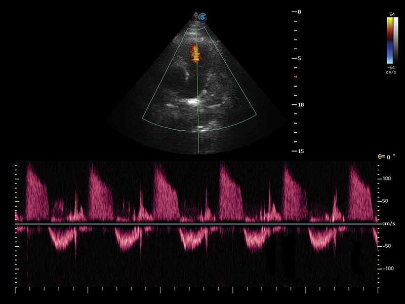
The 128-physical-channel architecture supports high-detail real-time visualization, with B-mode frame rates up to 1,493 fps and 10 imaging levels. The system supports B, 2B, 4B, M, CFM, PDI, DirPDI, PW, HPRF, CW, TDI, Elastography, and 3D/4D imaging modes, alongside Tissue Harmonic Imaging for improved contrast resolution. Continuous Wave (CW) Doppler supports high-velocity flow assessment, and Tissue Doppler Imaging (TDI) provides quantitative evaluation of local myocardial movements and functions.
C-xlasto Elastography and Contrast-Enhanced Imaging
C-xlasto elastography calculates Strain Ratio based on tissue stiffness variation, with a strain curve recording applied pressure to support lesion and tumor assessment in soft tissue. Contrast-enhanced imaging uses contrast agents to visualize small veins, organ perfusion, and blood flow with improved resolution and deeper penetration, maximizing the short duration of contrast bubbles for clearer organ imaging.
21.5-Inch HD LED Monitor and 14-Inch Touchscreen
The 21.5-inch HD LED monitor provides 1,920 × 1,080 pixel resolution for detailed image review, alongside a 14-inch touchscreen control panel for fast, direct operation. The fully adjustable control panel, integrated gel warmer, built-in workstation, and wireless Wi-Fi connection support ergonomic and efficient workflow across different scanning positions.
500 GB Storage and 200,000 Image Capacity
The system includes 500 GB hard-disk storage with capacity for 200,000 images and 1,000 cine loops in B-mode, supporting retrospective review and record retention. ECG access and foot-switch connectivity are supported as additional sockets for cardiac timing and hands-free operation.
Broad Clinical Applications
The FDA-cleared indications cover fetal, abdominal, pediatric, small-organ (breast, testes, thyroid), cephalic (neonatal and adult), transrectal, trans-vaginal, peripheral vascular, cerebral vascular, musculoskeletal (conventional and superficial), cardiac (pediatric and adult), trans-esophageal (cardiac), laparoscopic, OB/GYN, and urology applications. The 1–17 MHz frequency range and broad probe portfolio support shared-service imaging across obstetrics, gynecology, cardiology, radiology, and general practice.
Technical Specifications
| Specification | Detail |
|---|---|
| Device Type | Trolley-based premium color Doppler ultrasound system |
| Regulatory Status | FDA 510(k) K152854 (Jan 13, 2016) — S50 Digital Color Doppler Ultrasound System; Class II, Product Code IYN, 21 CFR 892.1550; Traditional review, 106 days; “No clinical data was required” per FDA summary; CE marking and ISO 13485 referenced in distributor documentation; no independent peer-reviewed clinical validation study specific to S50 identified |
| Physical Channels | 128 |
| Imaging Modes | B (including Tissue Harmonic Imaging), 2B, 4B, M, CFM, PDI, DirPDI, PW, HPRF, CW, TDI, Elastography, 3D/4D |
| Frequency Range | 1–17 MHz |
| Probe Compatibility | Up to 24 probe types; 6 sockets (4 active ports, 1 parking position, 1 pencil); HD probes with 256 elements |
| Probe Types | Linear, convex, single-crystal convex, micro-convex, endocavitary, phased array, volume, intraoperative, TEE, bi-plane, pencil, endocavitary 4D, laparoscope |
| Frame Rate (B-Mode) | Up to 1,493 fps |
| Display | 21.5-inch HD LED monitor, 1,920 × 1,080 pixels; 14-inch touchscreen control panel |
| Storage | 500 GB HDD; 200,000 images; 1,000 B-mode cine loops |
| Additional Sockets | ECG access, foot-switch access |
| Imaging Features | C-xlasto elastography (Strain Ratio, strain curve); Contrast-enhanced imaging; Tissue Doppler Imaging |
| Connectivity | Wireless Wi-Fi; built-in workstation |
| Ergonomics | Fully adjustable control panel, integrated gel warmer, front and rear handles |
| Clinical Applications | Fetal, Abdominal, Pediatric, Small Organ, Cephalic, Transrectal, Trans-vaginal, Peripheral Vascular, Cerebral Vascular, Musculoskeletal, Cardiac, Trans-esophageal, Laparoscopic, OB/GYN, Urology |
| Intended Use | General-purpose ultrasonic imaging for diagnostic evaluation of the clinical applications listed above by a qualified physician |
Why Choose the SonoScape S50?