

WhatsApp USA: +1 6032769350 | WhatsApp HK: +852 63697831
Global Healthcare Medical
SonoScape S80 Trolley Color Doppler Ultrasound System
SonoScape S80 Trolley Color Doppler Ultrasound System
The SonoScape S80 is a premium trolley-based color Doppler ultrasound system engineered for multi-modality clinical imaging across radiology, obstetrics and gynecology, musculoskeletal, and interventional applications. It is built on SonoScape's C-Field+™ architecture, a platform designed around precise signal-to-noise ratio, spatial and contrast resolution, and frame rate, supported by intelligent XPUs hybrid hardware and accelerated computing for boosted processing. The system is positioned as a flagship systemic-application platform, offering a comprehensive clinical solution that spans abdominal, OB/GYN, MSK, cardiac, and small-parts imaging within a single cart-based configuration.
The S80 is listed in the FDA Establishment Registration & Device Listing as the “S80 Digital Color Doppler Ultrasound System” , under Product Code IYN (System, Imaging, Pulsed Doppler, Ultrasonic), Class 2, Regulation 892.1550, with Premarket Submission Number K222596 cited in the listing. The K222596 clearance covers the S90 Exp Series, which includes the S80, S80 Elite, S80 Exp, and S80 Plus models. Distributor documentation also references CE MDR certification for the S80 platform. No independent peer-reviewed clinical validation study specific to the S80 was identified in the public literature during preparation of this page. Availability, labeling, and cleared claims vary by market.
The imaging chain is anchored by a premium transducer portfolio—convex, linear, phased array, volume, and specialty probes—with wideband frequency ranges designed to balance resolution and penetration for minute detail detection. Advanced hemodynamic tools include FHR for microvascular structure visualization, Bright Flow for 3D-like color Doppler flow without a volume transducer, and Micro F+ for separating subtle low-velocity flow signals from tissue motion using an innovative Matrix E adaptive filter algorithm.
The S80 supports department-specific clinical modules: S-MSK for automatic musculoskeletal joint section identification, S-Fetus® for one-click standard fetal plane acquisition and biometry, SWE for liver fibrosis quantification with a quality map, ATI for liver steatosis assessment through attenuation coefficient measurement, URI-CEUS for micron-scale angiogram reconstruction, Contrast HFR for high-frame-rate lesion perfusion display, SonoFusion for CT/MR–ultrasound fusion using a probe-integrated magnetic sensor, S-Thyroid with ACR TI-RADS-based classification, and S-Breast with BI-RADS standardized reporting.
Workflow features include Probe Auto Wakeup, SonoSynch for real-time remote consultation, SonoDrop for wireless image and report transfer, SonoAssistant for automated protocols, SonoHelp for on-screen tutorials, and SonoTransfer for Windows-based data sharing. The system is presented with a 24-inch LED monitor and 15-inch tilting touchscreen for high-definition viewing and streamlined operation.
Key Features & Benefits
C-Field+™ Architecture with Intelligent XPUs Hybrid Hardware
The C-Field+™ platform integrates precise field imaging transmission, adaptive aggregation reconstruction, and intelligent XPUs hybrid hardware to optimize signal-to-noise ratio, spatial and contrast resolution, and frame rate. This architecture is designed to deliver pure signal and refined imaging across different body habitus and clinical applications, within the physical limits of the installed probe set.
Premium Transducer Portfolio: Convex, Linear, Phased, Volume, Specialty
The S80 supports a broad range of transducers—convex array, linear array, phased array, volume probe, and specialty probes—with wideband frequency ranges that balance resolution and penetration for enhanced minute detail detection. The evolutional transducers are positioned for excellent 2D image clarity and color sensitivity, supporting confident assessment in anatomy and pathology.
Advanced Hemodynamics: FHR, Bright Flow, and Micro F+
FHR enables observation of microvascular structure within organs with enhanced sensitivity and improved resolution. Bright Flow provides 3D-like color Doppler flow visualization without a volume transducer, strengthening vessel wall boundary definition and reducing flow spillover. Micro F+ uses an innovative Matrix E adaptive filter algorithm to effectively filter out soft tissue and noise signals, retaining ultra-low-velocity fine blood flow signals and improving spatial resolution for tissue and lesion perfusion assessment. These are imaging capabilities—clinical value depends on proper probe selection and clinician review.
Department-Specific Clinical Modules: S-MSK, S-Fetus, SWE, ATI, and CEUS
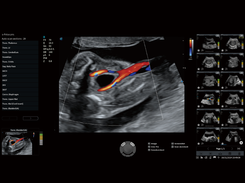
The S-MSK module automatically identifies standard musculoskeletal joint sections and highlights anatomical structures from the cine loop with one click. S-Fetus® intelligently identifies 29 standard fetal sections and automatically obtains 13 biometric measurements with a single click, supporting standardized and efficient prenatal assessment. SWE (Shear Wave Elastography) supports quantitative liver fibrosis assessment with a quality map for measurement reliability, including P-SWE and S-SWE methods. ATI (Acoustic Attenuation Imaging) quantifies liver steatosis through attenuation coefficient measurement. URI-CEUS reconstructs micron-scale angiograms with high-resolution morphological and hemodynamic information. Contrast HFR dynamically displays lesion perfusion at high frame rates, combined with MFI, MFI Time, and Mix mode for multi-perspective perfusion review.
SonoFusion with Probe-Integrated Magnetic Sensor
SonoFusion fuses ultrasound data with CT/MR data for simultaneous display by image registration, using a built-in magnetic sensor in the probe—eliminating the need for an external magnetic sensor. This supports comprehensive disease analysis where multimodal correlation is clinically relevant.
S-Breast and S-Thyroid with Standardized Classification Frameworks
S-Breast applies BI-RADS guidelines for standardized reporting and automatic lesion boundary extraction and classification. S-Thyroid supports detection and classification of suspicious thyroid lesions based on ACR TI-RADS guidelines, with automatic boundary definition and report generation. These tools are classification and reporting aids—they support the clinician's assessment and do not constitute a diagnosis on their own.
24-Inch LED Monitor and 15-Inch Tilting Touchscreen
The system features a 24-inch LED monitor and a 15-inch tilting touchscreen, providing high-definition viewing and a straightforward workflow. The ergonomically optimized design and remote access solutions support productivity in daily clinical operations.
Workflow Assistants: Probe Auto Wakeup, SonoSynch, SonoDrop, and SonoHelp
Probe Auto Wakeup uses an intelligent motion boost sensor in the transducer for auto activation with multi-adjustable sensitivity levels. SonoSynch enables real-time shared interface and camera for remote consultation and parameter adjustment across multiple devices. SonoDrop supports wireless onsite transfer of images, video, and PDF reports. SonoAssistant helps streamline workflow, increase standardization, and reduce keystrokes and exam time. SonoHelp provides on-screen tutorials with probe placement, anatomy, and reference images. SonoTransfer transfers PDF reports, images, and cine to a shared Windows PC folder.
Technical Specifications
| Specification | Detail |
|---|---|
| Device Type | Trolley-based premium color Doppler ultrasound system |
| Architecture | C-Field+™ with precise field imaging transmission, adaptive aggregation reconstruction, and intelligent XPUs hybrid hardware |
| Display | 24-inch LED monitor; 15-inch tilting touchscreen |
| Transducer Types | Convex, linear, phased array, volume, specialty; wideband frequency range |
| Imaging Modes | B/W, Color Doppler, Spectral Doppler, Elastography, Contrast-Enhanced Ultrasound |
| Hemodynamic Technologies | FHR, Bright Flow, Micro F+ |
| Clinical Modules | S-MSK, S-Fetus®, SWE (P-SWE/S-SWE), ATI, URI-CEUS, Contrast HFR, SonoFusion, S-Breast (BI-RADS), S-Thyroid (ACR TI-RADS) |
| Fusion Imaging | SonoFusion with probe-integrated magnetic sensor; CT/MR fusion |
| Workflow Features | Probe Auto Wakeup, SonoSynch, SonoDrop, SonoAssistant, SonoHelp, SonoTransfer |
| Regulatory Status | FDA Device Listing: “S80 Digital Color Doppler Ultrasound System”; Product Code IYN, Class 2, Regulation 892.1550; Premarket Submission K222596; CE MDR referenced in distributor documentation; no independent peer-reviewed clinical validation study specific to S80 identified; confirm local registration before purchase |
| Intended Use | Multi-modality ultrasound imaging, measurement, display, and analysis for clinical assessment across abdominal, OB/GYN, MSK, and related applications by qualified healthcare professionals |
Why Choose the SonoScape S80?
| Clinical or Operational Challenge | S80 Solution |
|---|---|
| Need a premium shared-service ultrasound across radiology, OB/GYN, MSK, and interventional applications | C-Field+™ architecture with multi-modality imaging and a broad transducer portfolio |
| Need high image clarity and hemodynamic sensitivity for challenging flow | FHR, Bright Flow, and Micro F+ for microvascular, 3D-like flow, and low-velocity fine-vessel visualization |
| Need quantitative tissue characterization for liver, thyroid, and breast assessment | SWE for liver fibrosis, ATI for liver steatosis, S-Breast (BI-RADS), S-Thyroid (ACR TI-RADS) |
| Need automated fetal and MSK standard plane acquisition | S-Fetus® (29 planes, 13 biometry); S-MSK (automatic joint section identification) |
| Need multimodal correlation with CT/MR | SonoFusion with probe-integrated magnetic sensor; no external sensor required |
| Need efficient workflow and remote collaboration tools | Probe Auto Wakeup, SonoSynch, SonoDrop, SonoAssistant, SonoHelp, SonoTransfer |
| Need high-frame-rate contrast perfusion display | Contrast HFR and URI-CEUS for dynamic lesion perfusion and micron-scale angiogram reconstruction |
| Need regulatory documentation for U.S. and EU markets | FDA Device Listing (IYN, K222596) and CE MDR referenced in distributor documentation; confirm specific model status for local market before purchase |